Ultrasound Imaging: Different Types and Their Uses in Medical Diagnosis
Whether you’re a medical professional, radiology technician, or someone curious about innovations in healthcare, this piece will give you an insightful look into ultrasound imaging-one of the most versatile tools in modern medicine.
For background on medical imaging, see X-Ray imaging guide.
We’ll explore its types, applications, transducer technology, benefits, limitations, and a glimpse into future advancements.
What Is Ultrasound Imaging?
Ultrasound imaging is a trusted, non-invasive diagnostic tool used to visualize structures inside the body.
It employs high-frequency sound waves, also known as ultrasound waves, which bounce off tissues and organs to create images. Unlike X-rays or CT scans, diagnostic ultrasound doesn’t use radiation, making it safer for routine examinations, particularly during pregnancy.
How Ultrasound Works?
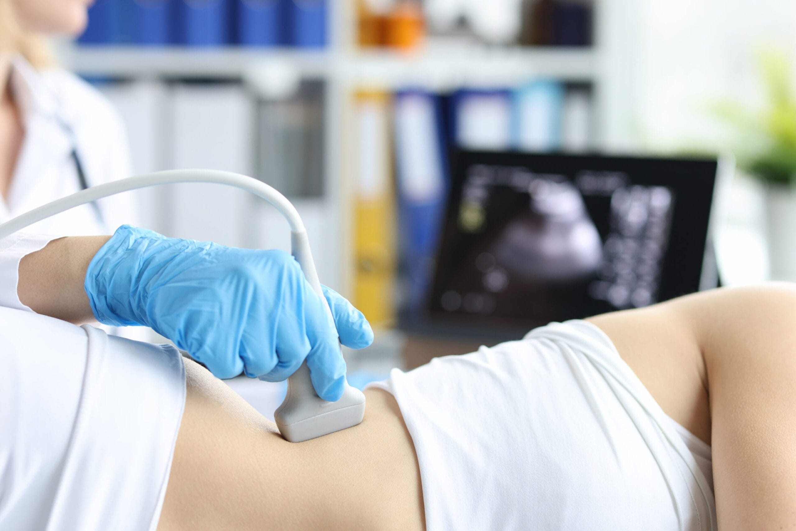
At the heart of every ultrasound machine is a transducer a device that emits and receives sound waves. These ultrasound waves reflect differently depending on the density of the structures they hit. The ultrasound machine then processes these waves into real-time images for medical imaging and diagnostic purposes.
A Brief History of Ultrasound in Medicine
Diagnostic ultrasound was first introduced into medicine during the 1950s. Initially, its applications were limited to simple observations, but advancements in technology have since transformed it into an indispensable medical imaging tool utilized across various medical fields.
Types of Ultrasound Imaging
Ultrasound is far more versatile than most people realize. Below, we’ll explore the key types and their uses in medical diagnosis.
Conventional Ultrasound

Conventional ultrasound uses sound waves to produce real-time images of internal organs and soft tissues.
2D Ultrasound
The most common type, 2D ultrasound produces flat images of internal structures. It’s primarily used for general diagnostics, such as examining organs or monitoring pregnancy.
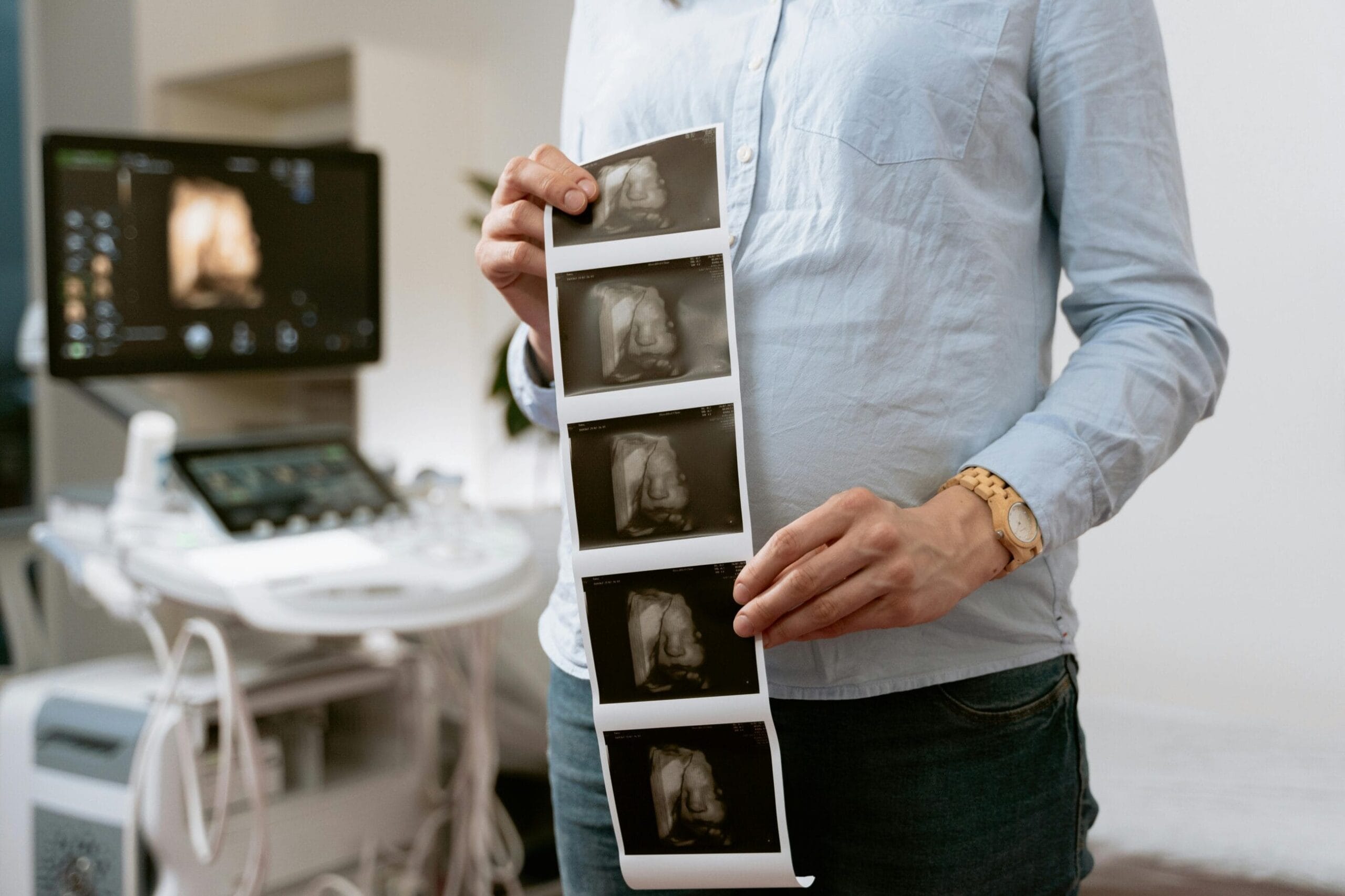
3D and 4D Ultrasound
While 3D ultrasound provides three-dimensional images, 4D ultrasound goes one step further with real-time imaging, often used in obstetrics to visualize fetal movements.
Specific Types and Their Applications
Ultrasound technology offers diverse applications, providing detailed insights into medical conditions with precision and reliability.
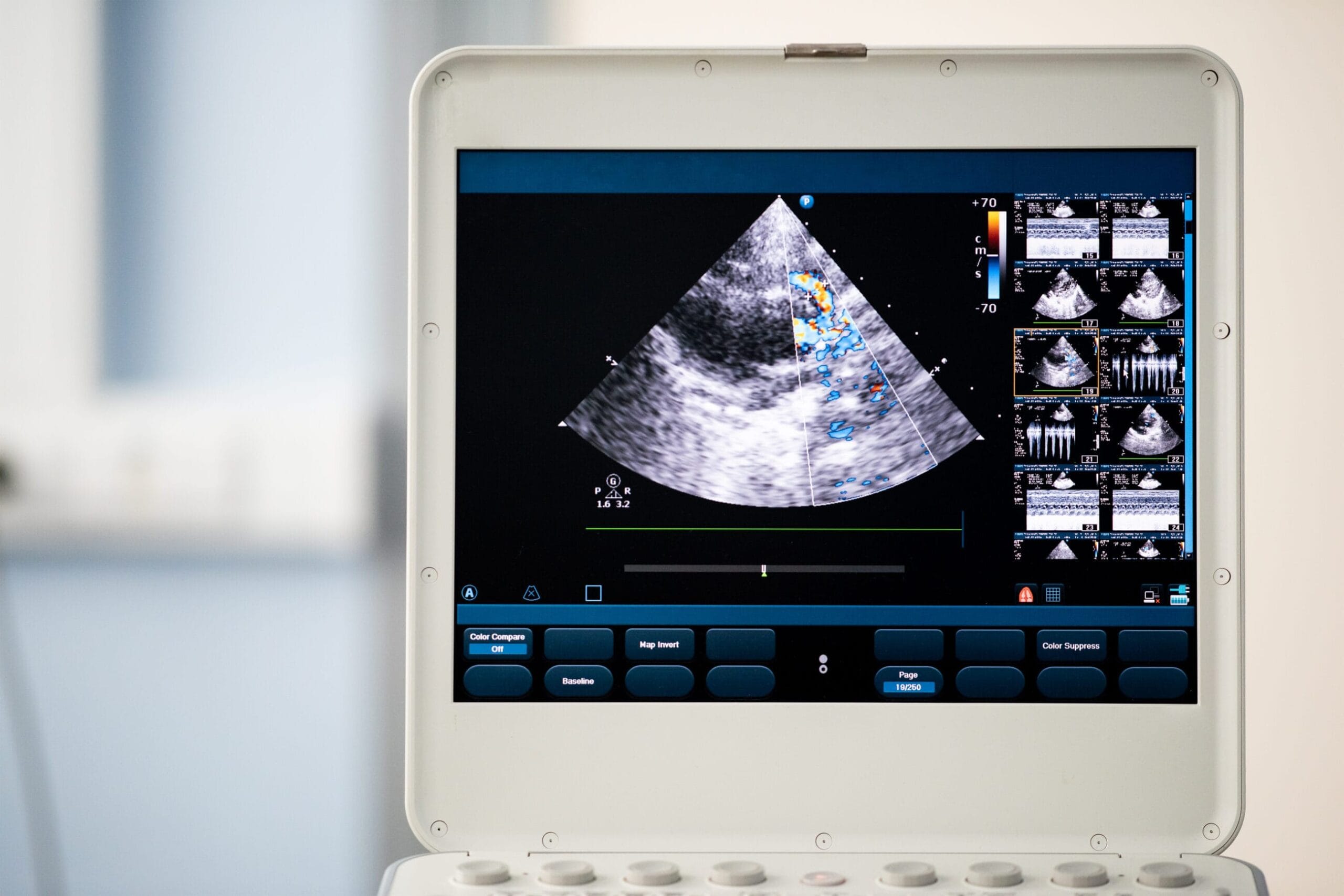
Doppler Ultrasound
Perfect for analyzing blood flow and circulation, Doppler techniques include:
- Color Doppler for visualizing flow direction and speed.
- Power Doppler for detecting slower blood flow.
- Spectral Doppler for measuring the blood’s velocity.
This type aids in identifying conditions such as blood clots or vessel blockages.
Transvaginal and Transrectal Ultrasound
Transvaginal ultrasound supports gynecological evaluations, and transrectal ultrasound is used for detailed imaging of the prostate gland, aiding in early detection of abnormalities.
Echocardiography
Used to assess heart health by imaging cardiac structures and blood flow, echocardiograms are essential for diagnosing heart diseases and monitoring treatments.
Endoscopic Ultrasound (EUS)
By integrating an ultrasound probe into an endoscope, healthcare providers achieve close-up views of internal organs via the digestive tract.
Point-of-Care Ultrasound (POCUS)
Portable and bedside-friendly, POCUS excels in emergency settings, aiding in quick diagnosis and guiding medical procedures.
Contrast-Enhanced Ultrasound (CEUS)
The addition of contrast agents to ultrasound enhances the clarity of images, making it ideal for examining organ abnormalities like liver lesions.
Intracavitary Ultrasound
Focused on imaging inside body cavities, this type is highly effective for gynecological or urological evaluations.
Application of Medical Ultrasound Imaging
Ultrasound has transformed how we diagnose and monitor health conditions. Here are some of its critical medical applications:
Obstetric and Prenatal Care
Ultrasound is integral to monitoring fetal development, evaluating risks, and ensuring maternal health throughout pregnancy.
Diagnostics for Abdominal Organs
From gallstones to liver fibrosis, ultrasound can detect numerous abdominal issues efficiently.
Cardiovascular Health
Ultrasound examination helps identify arterial blockages, monitor blood clots, and assess vascular conditions-all while being minimally invasive.
Musculoskeletal Imaging
It provides detailed images of tendons, muscles, and joints, proving particularly useful in sports medicine.
Cancer Detection and Biopsy Guidance
Ultrasound supports the early detection of abnormal growths and assists doctors in performing biopsies with precision.
Emergency Medicine
FAST (Focused Assessment with Sonography for Trauma) helps identify internal bleeding quickly in trauma settings.
Urology and Sexual Health
Conditions such as ovarian cysts, uterine fibroids, and prostate irregularities are often diagnosed using ultrasound.
Benefits of Ultrasound Imaging
Ultrasound is generally offers a wealth of benefits that make it a go-to imaging tool in many scenarios:
- Non-invasive and Safe: It poses no radiation risk.
- Real-time Results: Immediate visualization aids faster diagnosis.
- Cost-effective: Ultrasound is more affordable compared to MRI or CT scans.
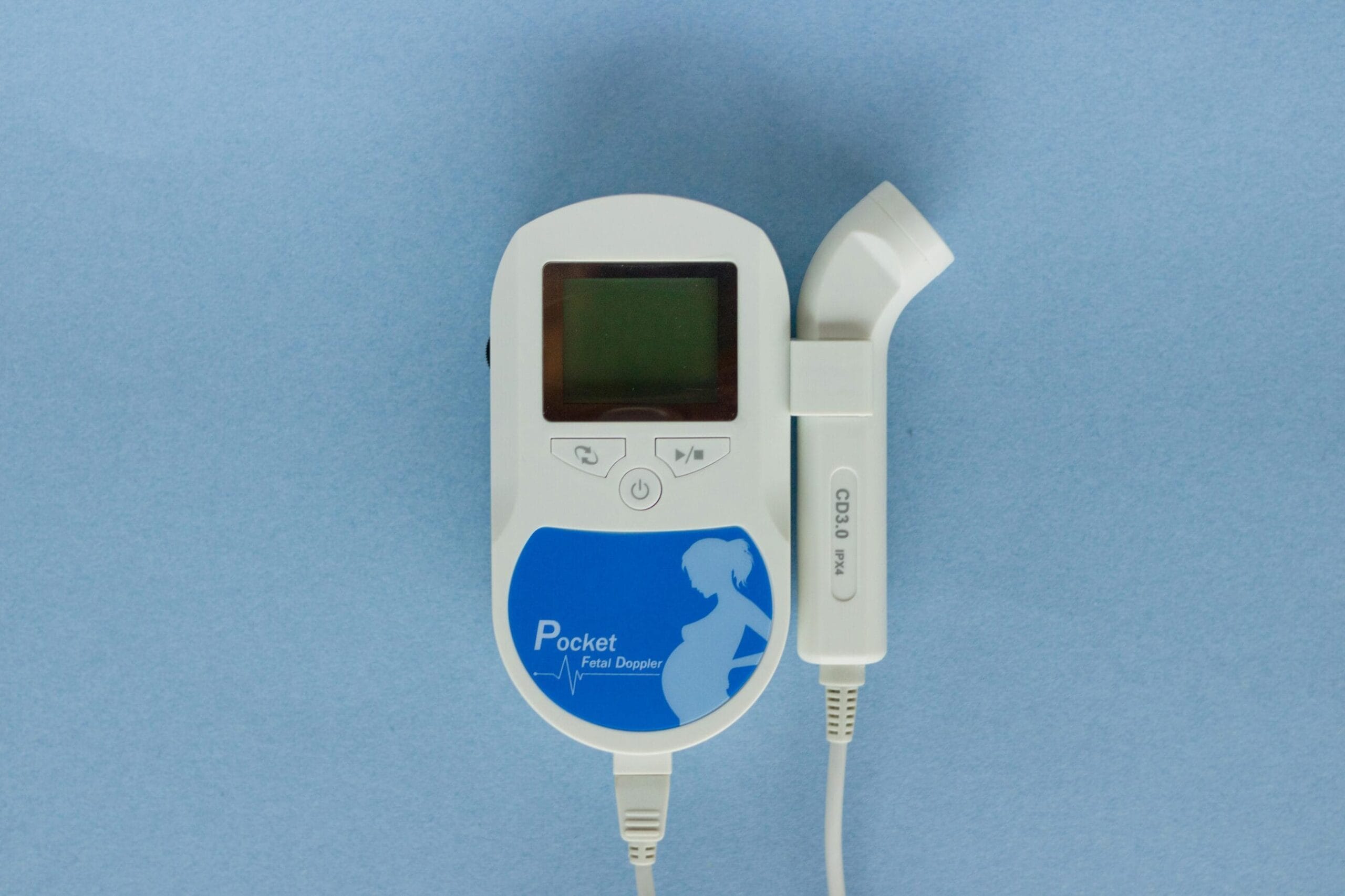
- Portable Technology: Thanks to miniaturization, portable ultrasound devices are now available, allowing care in remote locations.
- Versatility: It’s applicable in multiple medical fields, from obstetrics to emergency care.
Technological Advancements in Ultrasound
Advancements are continually shaping the future of ultrasound imaging:
- AI-Powered Ultrasound automates diagnostics, improves accuracy, and reduces human error.
- Miniaturization enables handheld devices for emergency or home care.
- Fusion Imaging integrates ultrasound with CT or MRI for more comprehensive evaluations.
- Wireless and Cloud Integration facilitates telemedicine and data sharing for remote consultations.
Limitations of Ultrasound Imaging
While ultrasound is versatile, it does have limitations:
- Limited Penetration Depth: It struggles to image deep tissues or organs in obese patients effectively.
- Operator Dependency: Image quality highly depends on the technician’s expertise.
- Reduced Resolution compared to advanced imaging tools like MRI or CT.
- Artefacts in Imaging can create challenges in interpreting results.
Ethical and Safety Considerations in Ultrasound
When used appropriately, ultrasound imaging is safe. However, overuse or non-medical applications-like gender reveals-should be avoided. Always ensure scans are guided by medical necessity and adhere to international guidelines.
Future Trends in Ultrasound Imaging
The future is bright for ultrasound technology:
- AI Integration will revolutionize diagnostics.
- Nanotechnology will enhance contrast agents for enhanced imaging.
- 5G Connectivity will expand tele-ultrasound services, increasing accessibility.
- Wearable Ultrasound for continuous monitoring is on the horizon, transforming how we approach health management.
Encouragement to Utilize Ultrasound for Preventive Care
Ultrasound imaging is more than a diagnostic tool-it’s a gateway to early detection and proactive health management. This technique uses gel applied to the skin to ensure clear imaging of internal structures, such as blood vessels, organs, and tissues. For residents of Singapore, it remains a highly accessible and cost-effective method for preventive care and is considered to be safe. Whether it’s a general health check-up or a targeted test like a prostate or pelvic scan, ultrasound keeps your health in focus.
At Mediway Medical, we’re committed to delivering high-quality care with modern diagnostic technology and experienced professionals. If you’re considering a health check-up or have questions about how ultrasound imaging could support your care, don’t hesitate to reach out.
Related Reading
Explore our X-ray imaging service.
Frequently Asked Questions
01 Is ultrasound imaging safe?
Yes, ultrasound imaging method is a non-invasive and safe imaging technique. It does not use ionizing radiation, making it a preferred option for many patients, including pregnant women and children.
02 What are the common uses of ultrasound?
Ultrasound is widely used for monitoring fetus during pregnancy, evaluating abdominal organs, diagnosing vascular conditions, guiding biopsies, and assessing musculoskeletal injuries.
03 Do I need to prepare for an ultrasound exam?
Preparation depends on the type of ultrasound. For abdominal scans, fasting may be required, while for pelvic or obstetric ultrasounds, drinking water to fill the bladder might be necessary. Your healthcare provider will give specific instructions.
04 How long does an ultrasound exam take?
Most ultrasound exams are quick and typically take 15 to 45 minutes, depending on the area being assessed and the complexity of the procedure.
05 What is the Difference Between Sonography and a CT Scan?
Sonography uses sound waves to create images, while a CT scan utilizes X-rays for detailed cross-sectional imaging. Sonography is non-invasive with no radiation exposure, unlike CT scans.


Why Choose Us
Choose Mediway Medical for reliable and efficient medical examinations, with on-site facilities, streamlined processes, and clear reporting timelines.
One-Stop Medical Centre
We provide comprehensive services in a single visit, including on-site chest X-ray and required medical tests.
Patient-Centred Experience
Our dedicated healthcare team focuses on providing clear guidance, professional care, and a comfortable clinic experience.
Minimum Waiting Time
We prioritise efficient workflows to keep waiting times short while maintaining accuracy and care quality.
Fast Turnaround Time
Medical reports are ready by the next working day, with same-day reporting available for selected cases.
Book Your Visit
Walk-ins welcome up to 30 minutes before closing.
Directions
Located in central Singapore, just a 2-minute walk from Clarke Quay MRT.
Mon–Fri before 5/6pm: $2.00 for 1st hr, $1.00 for next subsequent 30min from 7am to 5pm
Mon–Fri after 5/6pm: $3.21/entry from 5pm to 7am the following day
Sat: $2.00 for 1st hr, $1.00 for next subsequent 30min from 7am to 5pm, $3.21/entry from 5pm to 7am the following day
Sun/PH: $3.21/entry from 7am to 7am the following day
Exit E. 2-minute walk along the river towards The Riverwalk building. We are at #B1-26/29.